

Physicians
Orthopedic Surgery
Shoulder & Knee
Sports Medicine
 Healthgrades
Orthopedic Surgery
Shoulder & Knee
Sports Medicine
 Healthgrades
Orthopedic Surgery
Shoulder & Knee
Sports Medicine
 Healthgrades
Orthopedic Surgery
Shoulder & Knee
Sports Medicine
 Healthgrades
Orthopedic Surgery
Shoulder & Knee
Sports Medicine
 Healthgrades
Body parts
The anterior cruciate ligament (ACL) is found inside your knee and courses from thighbone (femur) to the shinbone (tibia). The ACL forms an “X” with the posterior cruciate ligament (PCL), hence the name “cruciate” from the Latin word meaning “cross”. The ACL and PCL control the forward (anterior) and backwards (posterior) stability of your knee. Specifically, the ACL prevents the shinbone from sliding out in front of (or anterior to) the thighbone. It also helps stabilize your knee when it rotates.
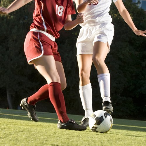
Injury to the ACL is common among football, basketball and soccer players but can occur in any sport. ACL injuries include sprains and stretches, partial tears and complete tears.
Suddenly stopping, changing direction, landing and twisting from a jump or slowing down too fast when running can damage the ACL. ACL injuries are especially common among athletes who participate in sports such as soccer, football and basketball because these sports require quick-adjustment movements. Think of the ACL as a flexible fence guarding knee joint. The forceful pressure of sudden pivoting and shifting direction can break through the fence.
Due to differences in pelvis and leg alignment and ligament tension, female athletes are more susceptible to ACL injuries than men.
For a diagnosis and treatment plan, your Orthopedic Institute of New Jersey (OINJ) knee doctor will take a medical history, determine when the injury occurred and do a physical exam by comparing your injured knee to your healthy knee. Your knee specialist may order an X-ray, as well as an MRI to confirm diagnosis, rule out fracture and evaluate the degree of ligament, meniscus (cartilage in the knee that acts as a shock absorber) and other cartilage involvement.
Imaging can also help your knee doctor determine the extent of the damage, which can range from a simple sprain or stretch of the ACL to a full tear, which completely destabilizes the knee.
Treatment options correspond to the degree of the injury and your level of activity, with the goal of stabilizing and strengthening your knee so it can bear the level of activity that will be required of it. A tear won’t heal itself without surgery, but rest, bracing and physical therapy will be all that’s needed for a non-athlete with a minor ACL sprain.
If surgery is needed for your ACL injury, your ACL surgeon will reconstruct the ACL with graft, either using your own tissue or cadaver tissue. For young athletes, using your own tissue (autograft) is preferable as it leads to lower rates of ACL retear. Autograft options include hamstring tendon or patellar tendon with bone attached. A torn ACL is replaced rather than repaired.
During surgery, your knee surgeon will reconstruct your ligament with minimally invasive techniques that result in less pain and shorter recovery time. You will start gentle range of motion exercises immediately following surgery and begin physical therapy shortly after. Physical therapy continues for approximately 6 months, and includes home exercises to optimize your recovery and outcome.
Restoring knee stability is ultimately the name of the game. But healing is a process. For many athletes, it can take up to a year of playing before they feel confident going out and doing what they did before the injury.